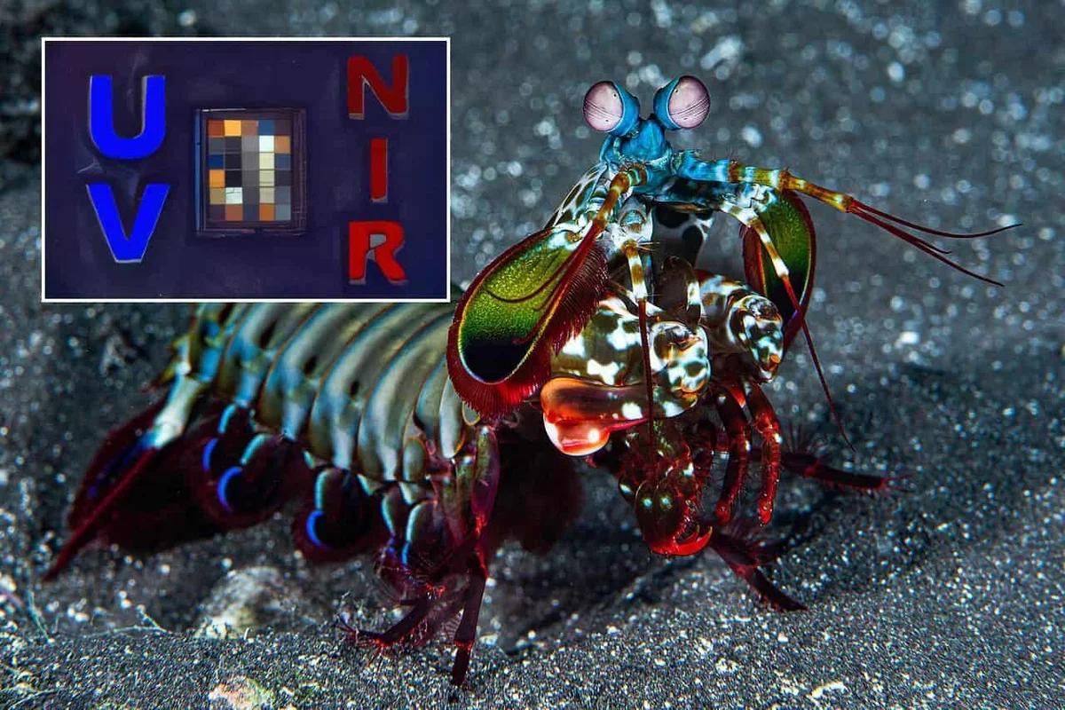
The mantis shrimp, a fascinating marine creature, is renowned for its extraordinary vision. Researchers have now harnessed this natural marvel to create a groundbreaking camera designed for use in surgical settings.
This innovative device replicates the shrimp's ability to perceive a wide spectrum of light, including ultraviolet, visible, and near-infrared wavelengths, all captured on a compact chip. This technology aims to assist surgeons in identifying not only the location of lymph nodes but also whether they contain hidden cancer cells during operations.
Enhancing Surgical Precision
During breast cancer surgery, surgeons face the critical task of removing the primary tumor while assessing the lymphatic system, which can harbor cancer cells. Lymph nodes serve as filters, trapping these rogue cells before they can spread further. The challenge lies in determining which nodes to biopsy or remove, balancing the risk of cancer spread against potential complications like lymphedema from excessive removal.
Current methods often rely on chemical dyes that provide limited information about the cancer's status, leaving surgeons to make decisions without immediate insights into the tissue pathology.
Viktor Gruev from the University of Illinois at Urbana-Champaign emphasized the limitations of existing tools, stating, "They can show where lymph is draining but can't reliably indicate whether a particular lymph node is involved with cancer while the operation is underway." This gap can lead to either overtreatment or undertreatment, necessitating further procedures.
The Science Behind the Camera
The camera's design draws inspiration from the mantis shrimp's unique photoreceptors, which allow it to see beyond the human visible spectrum. By integrating pixel-level filters and stacked light-sensing layers, the new camera can capture different light types simultaneously. This technology enables surgeons to receive real-time information about lymph nodes during surgery.
Instead of traditional glass lenses, a specialized mirror-based lens system maintains focus across various wavelengths, while custom software reconstructs the images into a coherent view. "This allows our camera to collect several kinds of optical information from exactly the same place at the same time," Gruev noted.
Spotting Cancerous Tissue
The camera utilizes a standard fluorescent dye, indocyanine green (ICG), which highlights lymph nodes under near-infrared light. Once identified, the camera employs its ultraviolet capabilities to differentiate between healthy and cancerous tissues based on their fluorescence. This method does not require additional cancer-targeting chemicals, providing a significant advantage in surgical settings.
In tests involving 94 lymph nodes from breast cancer patients, the camera demonstrated impressive accuracy, identifying cancerous nodes with 97% sensitivity and 89% specificity. The next steps involve extensive testing to ensure reliability across diverse patient groups and refining the system for practical use in operating rooms.
If successful, this technology could revolutionize cancer surgery, enhancing precision and potentially improving outcomes not only for breast cancer patients but for various cancers where lymph node assessment is crucial.